About Paper
Venue / Year: MICCAI 2015 Link: https://arxiv.org/abs/1505.04597
TL;DR
- Problem: Extremely small dataset sizes for biomedical image segmentation (lack of annotated data).
- Method: A U-shaped encoder-decoder architecture with skip connections to combine global context and precise localization, heavily relying on strong data augmentation.
- Result: Achieves state-of-the-art results trained end-to-end from very few images; won ISBI challenges by a large margin; extremely fast inference (<1s per image).
Background & Motivation
- Why is this research needed? (Why now?)
- Deep learning typically requires thousands of annotated samples, but annotating biomedical images is expensive, time-consuming, and requires domain experts.
- Drawbacks of existing methods:
- Sliding-window CNNs: Process each patch individually, making it slow and computationally redundant due to overlapping patches.
- Trade-off between patch size and accuracy: Larger patches capture more context but require more max-pooling layers, which discard local spatial information. Thus, there's a trade-off between speed, context, and localization accuracy.
- Classification-based approaches: Struggle to balance global context capture with precise pixel-level localization.
Core Idea / Method
- U-Net Architecture (Encoder-Decoder):
- Contracting Path (Encoder): Repeated Conv + Max Pooling to capture broad context and abstract features.
- Expanding Path (Decoder): Up-convolutions (transposed convolutions) to progressively restore spatial resolution for precise localization.
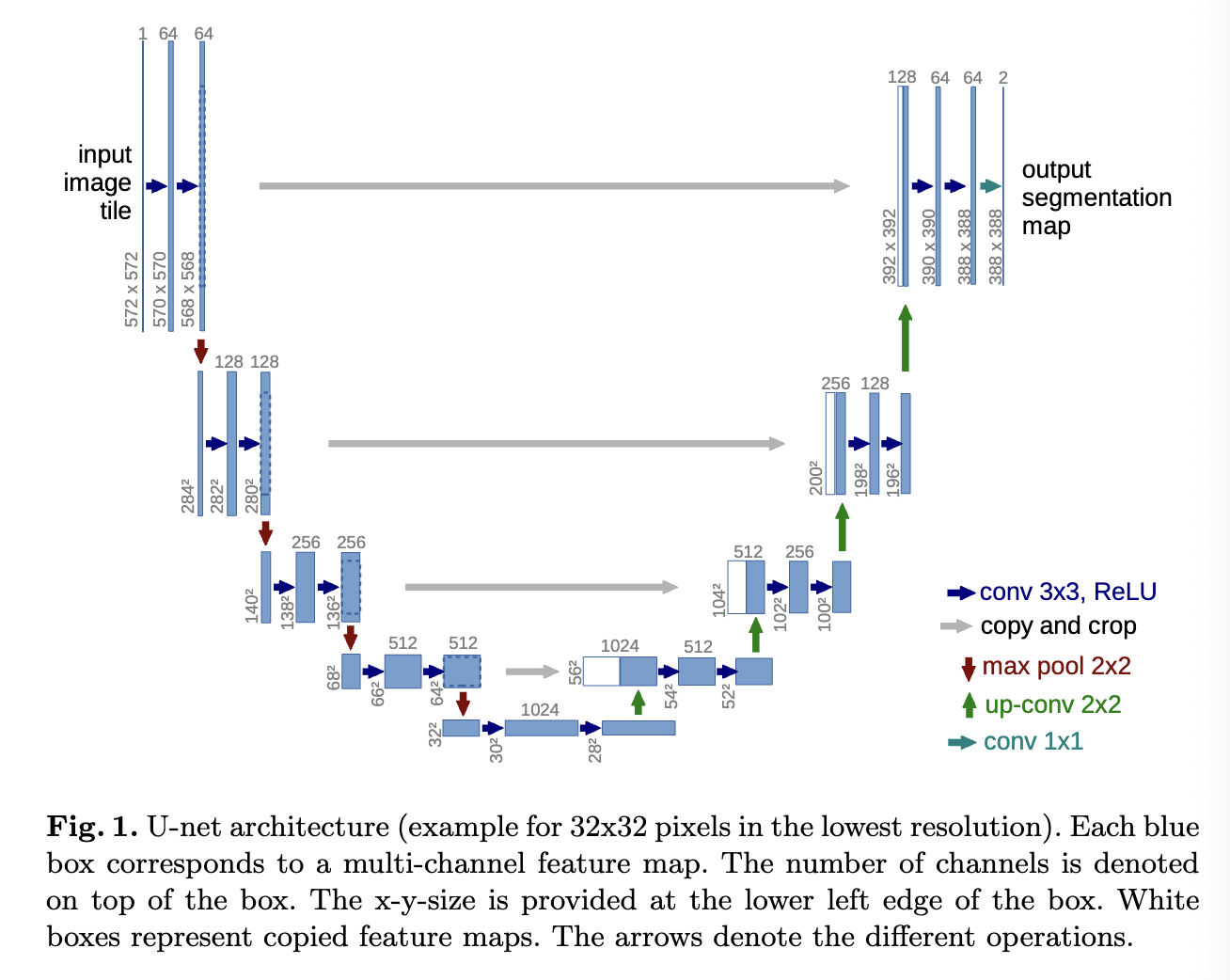
- Skip Connections: Concatenating high-resolution feature maps from the encoder to the corresponding decoder layers. This compensates for the spatial details (edges, shapes) lost during pooling.
- Strong Data Augmentation: Heavy use of elastic deformations to teach the network invariance to such variations, which is the key to succeeding with very little data.
- Overlap-tile strategy: Predicting larger tiles and cropping the center to ensure valid, seamless predictions at image borders without boundary artifacts.
- Weighted Loss Map: Assigning higher weights to pixels at the boundaries between touching cells to force the network to separate them.
Experiments & Results
- ISBI 2012 (Segmentation of neuronal structures in EM stacks): Outperformed prior sliding-window CNNs (evaluated via Warping Error, etc.).
- ISBI Cell Tracking Challenge 2015 (Phase contrast and DIC images): Won by a large margin.
- Speed: Segmentation of a 512x512 image takes less than a second on a modern GPU.
- Data Efficiency: Demonstrated that highly accurate models can be trained end-to-end with just a few dozen annotated images.
Pros & Cons
- Pros:
- Exceptional performance even with very limited training data.
- Skip connections yield extremely sharp and precise object boundaries.
- End-to-end trainable and incredibly fast at inference.
- Simple, elegant, and highly versatile architecture; became the de facto standard for segmentation.
- Cons:
- High memory consumption due to storing multiple high-resolution feature maps for skip connections (especially problematic for 3D U-Net or high-res inputs).
- Being CNN-based, it has a limited receptive field compared to modern Vision Transformers, making it less effective at capturing extremely long-range global dependencies.
My Takeaway
- The elegant fusion of "context acquisition (downsampling)" and "precise localization (upsampling)" via skip connections is a masterclass in architecture design.
- A historic milestone that transcended biomedical imaging, heavily influencing general segmentation tasks and even modern generative models (e.g., the U-Net noise predictor in Stable Diffusion).
- A powerful reminder of the importance of domain-specific data augmentation (like elastic deformations) and architectural ingenuity when applying AI to data-scarce domains.